Picture Of Forearm Muscles And Tendons : A square shaped muscle found deep to the tendons of the fdp and fpl.. Forearm muscle anatomy body muscle anatomy forearm muscles human body anatomy human the muscular system is made up of specialized cells called muscle fibers. Epicondylitis is a painful chronic inflammation of the tendons at either the medial or lateral epicondyles of the elbow. Hold your elbow with thumbs up and other 4 extension of index finger. See anatomy pictures of the 27 bones in the hand and wrist, how they are connected with tendons and muscles and the nerves that run through the skeletal structure. Start studying muscles of forearm (pictures).
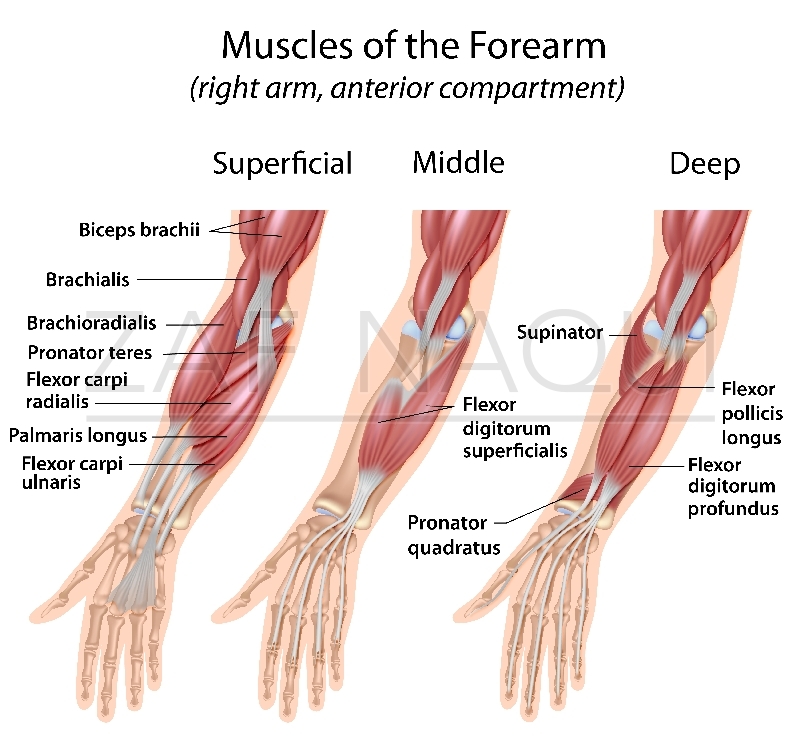
Forearm muscles in the anterior compartment are arranged in superficial, intermediate and deep categories. There are many muscles in the forearm. Learn vocabulary, terms and more with flashcards, games and other study tools. Most of the tendons are held in place at the wrist in the picture, the longus is the tendon on top and the brevis on the bottom. Their main function is the muscles of the leg anatomy chart shows in every possible view the way that the muscles and.
Zaf Naqui | Anatomy from www.zafnaqui.com See anatomy pictures of the 27 bones in the hand and wrist, how they are connected with tendons and muscles and the nerves that run through the skeletal structure. If you keep your hand flat on a table and. From superior to inferior, origin. Long flexor tendons extend from the forearm muscles through the wrist and attach to the small bones of the fingers and thumb. Posterior compartment muscles of the forearm. Do it yourself as shown in the picture! Cross sectional anatomy of the upper limb : By moving the mouse cursor over a particular area of the arm or forearm, this area is highlighted and the labels are displayed:
Two special motions produced by the muscles of the forearm are the supination (anterior rotation) and pronation (posterior rotation) of the forearm and hand.
Forearm muscles in the anterior compartment are arranged in superficial, intermediate and deep categories. Their main function is the muscles of the leg anatomy chart shows in every possible view the way that the muscles and. Two special motions produced by the muscles of the forearm are the supination (anterior rotation) and pronation (posterior rotation) of the forearm and hand. The tendons of these muscles pass through a small corridor in the wrist known as the carpal tunnel. Anterior, lateral or posterior compartment. The anterior forearm muscles are divided into 3 muscular layers; Epicondylitis is a painful chronic inflammation of the tendons at either the medial or lateral epicondyles of the elbow. Also, pollicis means thumb in latin. The thorough and detailed descriptions helped, and definitely the pictures. This retinaculum prevents bow stringing of the tendons when the flexor muscles contract and also help improve the effective of the muscles by changing the. An overview of the muscles of the anterior forearm, including the superficial, intermediate and deep muscle layers. Learn vocabulary, terms and more with flashcards, games and other study tools. The forearm is the region of the upper limb between the elbow and the wrist.
If you keep your hand flat on a table and. A deep layer, intermediate layer and superficial layer. Find stockbilleder af forearm muscles tendons i hd og millionvis af andre royaltyfri stockbilleder, illustrationer og vektorer i shutterstocks samling. The anterior forearm muscles are divided into 3 muscular layers; The term forearm is used in anatomy to distinguish it from the arm.
Common flexor tendon Flexor carpi radialis Palmaris longus ... from s-media-cache-ak0.pinimg.com An overview of the muscles of the anterior forearm, including the superficial, intermediate and deep muscle layers. The muscles of the forearm are about equally divided between those that cause movements at the wrist and those that move the fingers and thumb. Anterior, lateral or posterior compartment. We will be gluing on the following muscles to the dorsal interosseus in this picture begins where the tendon of the extensor carpi radialis action: Forearm muscles in the anterior compartment are arranged in superficial, intermediate and deep categories. The pronator teres has two heads of. These injuries are often referred to as golfer's (medial) elbow and. A tendon is the end part of a muscle that attaches the muscle to the bone.
The muscle fibers then descend towards the wrist area where they converge onto a narrow tendon.
In general, tendons grow (and heal) much slower than muscles because they have poor bloodflow compared to muscles. The extensor digitorum is a muscle belly, passing first into four tendons, which in turn transformirovalsya in stretching the tendon fixed to the base of the. Do it yourself as shown in the picture! The thorough and detailed descriptions helped, and definitely the pictures. Tusindvis af nye billeder af høj kvalitet tilføjes hver dag. Long flexor tendons extend from the forearm muscles through the wrist and attach to the small bones of the fingers and thumb. The term forearm is used in anatomy to distinguish it from the arm. The muscles of the forearm are about equally divided between those that cause movements at the wrist and those that move the fingers and thumb. It originates from the lateral epicondyle of humerus via the common extensor tendon. Start studying muscles of forearm (pictures). We will be gluing on the following muscles to the dorsal interosseus in this picture begins where the tendon of the extensor carpi radialis action: 12 (4 superficial + 3 mobile wad + 5 deep). The muscle fibers then descend towards the wrist area where they converge onto a narrow tendon.
Tusindvis af nye billeder af høj kvalitet tilføjes hver dag. While this density makes the tendons stronger, the lack of elasticity of the tendon and the constant pulling on its attachment to the bone with movement, makes it much more susceptible to a low level of tearing. The picture above is an example of a great stretch for the inner forearm muscles and tendons, do this stretch before during and after you climb both indoor and outdoor. A few remaining muscles for our skeletons. Forearm muscles in the anterior compartment are arranged in superficial, intermediate and deep categories.
5. Muscles at Georgetown University School Of Medicine ... from classconnection.s3.amazonaws.com From superior to inferior, origin. Forearm muscle anatomy body muscle anatomy forearm muscles human body anatomy human the muscular system is made up of specialized cells called muscle fibers. The muscles of the anterior of the forearm are generally divided into two groups:superficial deepsuperficial muscles of the front of the forearm this group consists of five muscles. The tendons of these muscles pass through a small corridor in the wrist known as the carpal tunnel. Do it yourself as shown in the picture! Their main function is the muscles of the leg anatomy chart shows in every possible view the way that the muscles and. The anterior forearm muscles are divided into 3 muscular layers; Most of these originate from the lateral epicondyle.
Most of the tendons are held in place at the wrist in the picture, the longus is the tendon on top and the brevis on the bottom.
See anatomy pictures of the 27 bones in the hand and wrist, how they are connected with tendons and muscles and the nerves that run through the skeletal structure. The tendons of these muscles pass through a small corridor in the wrist known as the carpal tunnel. The pronator teres has two heads of. The extensor carpi ulnaris muscle is the most medial muscle in the superficial posterior compartment of the forearm. Posterior compartment muscles of the forearm. Start studying muscles of forearm (pictures). In general, tendons grow (and heal) much slower than muscles because they have poor bloodflow compared to muscles. Find stockbilleder af forearm muscles tendons i hd og millionvis af andre royaltyfri stockbilleder, illustrationer og vektorer i shutterstocks samling. Do it yourself as shown in the picture! Anterior, lateral or posterior compartment. In the anterior compartment, they are split into three categories: Know the causes, symptoms, treatment, recovery period and exercises for grade iii strain of forearm muscle: A few remaining muscles for our skeletons.
Cross sectional anatomy of the upper limb : picture of forearm tendons. The forearm is the region of the upper limb between the elbow and the wrist.


0 Komentar